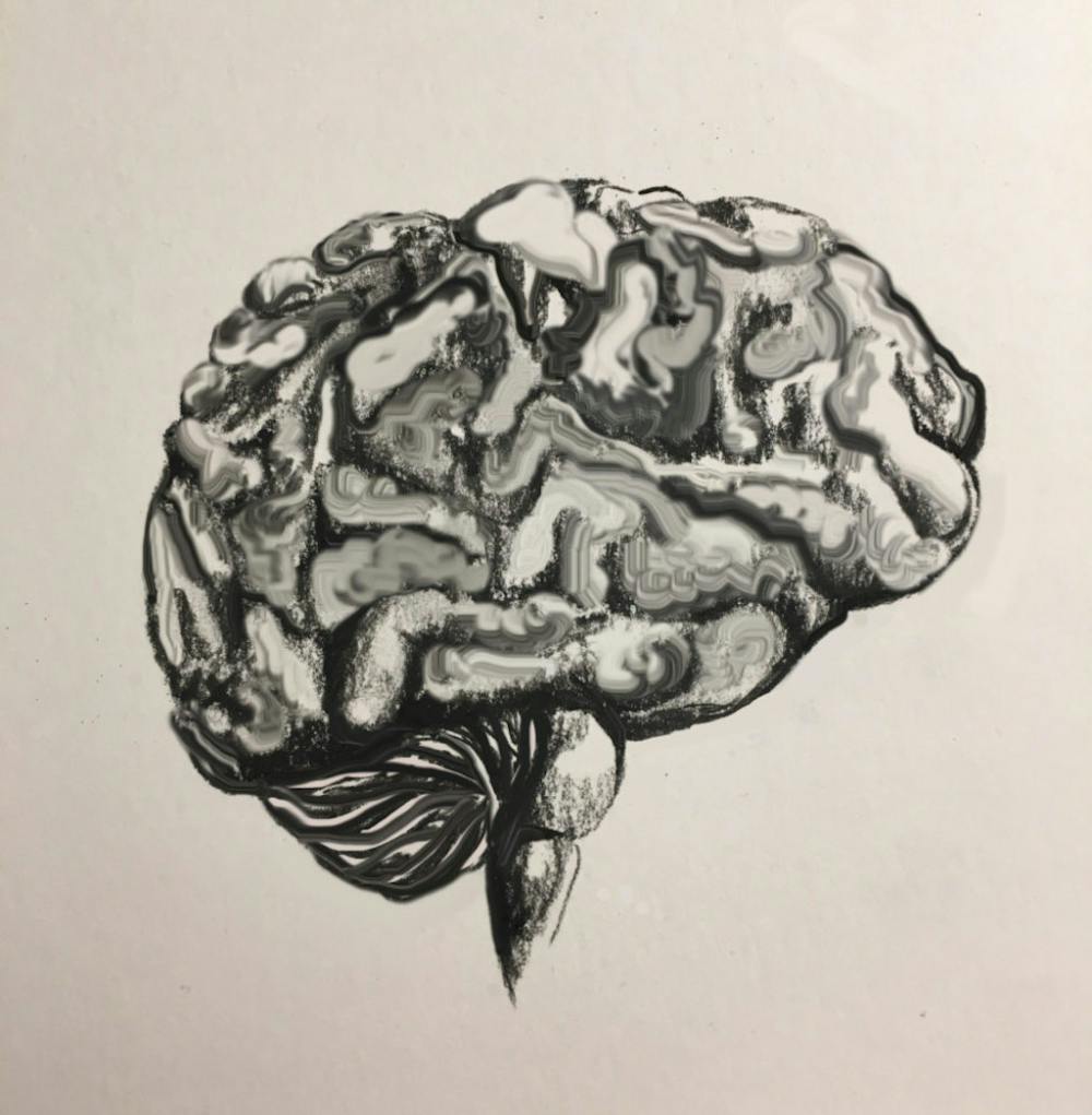
University researchers collaborated with the BrainGate clinical trial to identify the role of the middle frontal gyrus — an area in the front region of the brain — in planning movements based on auditory stimuli, including words and other sounds, through observations of a single participant in the trial.
The researchers involved in the BrainGate clinical trial hope to turn the knowledge they gain from their studies “into ever-better neuroengineering methods to allow people with paralysis to regain intuitive, reliable, dexterous control over communication devices such as tablet computers and, ultimately, one’s own limbs,” wrote Leigh Hochberg ’90, the director of the study, member of the BrainGate consortium leadership team and professor of engineering at Brown, in an email to The Herald.
The middle frontal gyrus is thought to be involved with executive behavior, including higher levels of cognition, decision-making and attention, said Zulfi Haneef, assistant professor of neurology at Baylor University and director of neurophysiology at the Michael E. DeBakey Veterans Affairs Medical Center, who is unaffiliated with the study.
But scientists “don't really know much about what exactly this part of the brain does,” Haneef said. The recent study published in Scientific Reports aims to help fill this gap in knowledge.
The trial recruited its participant through the BrainGate research consortium, which includes researchers from institutions such as Brown, Massachusetts General Hospital and Stanford University. Neuroscientists, computer scientists, engineers and clinicians across other institutions also contributed to the findings.
“One of the many benefits of the consortium is being able to constantly learn from each other and most importantly from our incredible clinical trial participants,” Hochberg wrote.
To obtain data from the participant, the researchers measured the activity of the middle frontal gyrus, which they believe is involved with the preparation and execution of movement. They implanted a grid of microelectrodes — about the size of a baby aspirin — into the brain tissue of the trial participant to record this activity, said Carlos Vargas-Irwin ’02 PhD’10, the study’s co-senior author and assistant professor of neuroscience research at Brown. The microelectrode array is a brain-computer interface technology that measures neural signals from the brain.
The trial participant, who is paralyzed at the arms and legs due to a spinal cord injury, was presented with visual cues and had to respond by moving a cursor toward the target stimuli that matched the description from the audio cues, according to a University press release. Since the participant was unable to physically move, his intended movements — thoughts in the form of neural activity detected by the implant — were decoded by computer software and used to control the motion of a cursor.
The microelectrodes recorded increased activity in the middle frontal gyrus in response to the audio cues preceding the movements.
The participant also underwent a passive listening control experiment in which he heard various sounds — including words used in the earlier part of the trial — but was not explicitly instructed to perform the movement task associated with that sound, Vargas-Irwin said. For example, the participant was originally told to attempt to move toward a red shape on the computer screen; but in this control experiment, he would hear “red” without having to respond to the verbal cue with a movement.
The researchers found that the middle frontal gyrus was still active in this control experiment, but not to the same extent as when accompanied by a command to move.
Certain disorders of the nervous system disrupt the pathways from the brain to muscles that normally carry signals commanding movement. As a result, the areas “in the brain that are responsible for controlling movement are still working, but the signal that they send to the muscles is interrupted,” Vargas-Irwin said. “The idea with (the brain-computer interface) is to tap into the movement intention directly and into (areas of the brain such as the middle frontal gyrus) that drive movement to be able to take those signals and do something useful with them.”
Moving forward, the researchers are interested to further examine if words with intrinsic meaning versus non-word sounds, such as high-pitched and low-pitched tones, impact brain activity in different ways.
Other researchers not affiliated with the study agree that this experiment would be a valuable future direction. Haneef said that due to the middle frontal gyrus’s proximity to an area of the brain associated with language, he “wouldn't be surprised if ... the middle frontal gyrus would have some connection to language.”
The researchers also hope to use these findings to assist in creating new devices for people that have sensory limitations and are more reliant on auditory cues than visual cues to perform daily activities.
“As we continue to have the extraordinary privilege to work closely with our clinical trial participants to develop technologies that will restore communication and mobility, a key opportunity is to learn as much about the human brain as possible,” Hochberg wrote.
“There’s still a lot to explore with the middle frontal gyrus and sound processing,” Vargas-Irwin said. “Together, I feel we really are pushing the frontiers of what’s known.”
A previous version of this article stated that Leigh Hochberg was MD’90. In fact, Hochberg received a Sc.B. from Brown. The Herald regrets the error.
Gabriella was a Science & Research editor at The Herald.


