Since the University’s MRI Research Facility opened for operations in early 2007 as part of the Sidney Frank Hall for Life Sciences, it has been a crucial resource for Brown researchers.
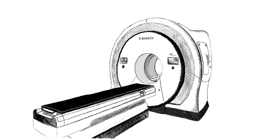
Magnetic Resonance Imaging has been a powerful and popular scanning technology critical to medical and research settings since its invention in the 1970s. Using magnets and radio waves, the tube-shaped MRI scanner can produce clear images showing organs, muscles or even blood vessels.
MRI scanners are expensive but very capable tools. Scanners themselves cost around $1 million per Tesla of force — the MRF machine has three Teslas.
The MRI room is also magnetically shielded, preventing the scanner’s magnetic field from extending beyond the facility and, according to MRF Associate Director of Research Michael Worden, potentially wiping passersby’s credit cards.
According to Worden, about 50 investigators from fields including neuroscience, engineering, chemistry and medicine are using the MRF. Most researchers work through Brown and Brown-affiliated Lifespan hospitals, but others come from other institutions like the University of Rhode Island, Worden wrote.
Theresa Desrochers, assistant professor of brain science and psychiatry and human behavior, studies cognitive and behavioral sequences in humans and animals. MRI is useful because it can help identify which parts of the brain are used and how those parts are engaged, Desrochers wrote in an email to The Herald.
“These kinds of sequences are like making breakfast or a cup of coffee,” she wrote. “You have to keep track of a series of steps and do them in a particular order. We are interested in how the brain does this tracking.”
Functional MRI, which can record activity over time in different parts of the brain, is particularly useful in Desrochers’s research because it can use similar techniques on both humans and animals.
For Samantha Buyungo ’24, an undergraduate research assistant in the Desrochers Lab, her first experience with MRI wasn’t in a research setting but a medical one after a knee injury between eighth and ninth grade.
“After getting an MRI, years later, to be working with the MRI (studying) sequential processing is interesting,” Buyungo said.
For her undergraduate thesis, Buyungo is comparing individuals with and without OCD in sequential processing tasks.
Buyongo said that in her present research, she aims to use MRI to more clearly visualize what is happening inside the brain — and hopes to continue doing so in the future.
Buyungo said MRI can show that mental health — along with mental illness — is a “concrete thing,” even if it isn’t immediately visible like physical health indicators.
“No matter how difficult my research gets, that’s something that I always like to come back to,” she said. “These are things that matter and can help you better understand someone else.”
Haley Keglovits GS, a PhD candidate, is studying how humans carry out executive functions in the brain in the lab of Professor of Cognitive, Linguistic and Psychological Sciences David Badre, who also chairs the department.
Executive functions are higher-level cognitive skills that allow individuals “to be flexible and not perform the same behaviors in response to a specific environmental cue,” Keglovits wrote in an email to The Herald. “For example, you might pick up your phone if you hear it beep when you are sitting at home, but not when you are driving.”
Keglovits’s study also employs functional MRI: Participants perform tasks related to executive brain functioning, and her team analyzes their patterns of brain activity and how the patterns change when participants encounter different objectives.
Scan results are “very different from what you might see on TV, where characters have (colored) dots popping up on their brain pictures in real-time,” Keglovits wrote. Instead, data processing is required before the researchers gather insights from a scan.
MRI research can create challenges, Keglovits and Desrochers wrote. For example, people must remain still in the scanner to prevent a blurry image — but keeping them comfortable might mean putting them to sleep, Keglovits added.
“If we make people too comfy and have them lay down in a dark room with pillows and a blanket, they can fall asleep while trying to do a task,” she wrote.
The backbone of the MRF’s research is a closely integrated team allowing researchers to turn MRI imaging into “robust and interpretable results,” Worden said. The MRF also relies on collaborations, extensive and rigorous safety protocols and the support of the Carney Institute for Brain Science, Worden said.
“The human brain is the most complicated thing in the known universe,” Worden said. “Understanding how the brain works, how cognition, behavior, disorders are related — these are incredibly important scientific endeavors.”

Ranjana “Jaanu” Ramesh is a Bruno Brief-er, photographer and Senior Staff Writer covering science & research. She loves service, empathetic medicine and working with kids. When not writing or studying comp neuro, Jaanu is outside, reading, skiing, or observing Providence wildlife (ie: squirrels).


