University researchers have developed a new connective tissue model to study disorders like fibrosis, a type of scarring that occurs when damage to the body causes normal tissue to be replaced by hard, fibrous connective tissue. In a paper recently published in the journal Advanced Science, the researchers demonstrate how this model can be used to better understand connective tissue mechanics and test potential therapies for connective tissue disorders.
“It's a new tool in the toolbox to understand fibrosis and to test potential drugs that could treat fibrosis, (which is) an unmet medical need,” said Jeffrey Morgan, professor of pathology and laboratory medicine and engineering.
Morgan is the director of the University's Center for Alternatives to Animals in Testing, an organization dedicated to developing replacements for animal models in research, according to the center’s website. His lab previously invented a method of cell culture in which cells grow in agarose gel molds rather than on polymer or protein scaffolds. Because of the properties of the gel mold, the cells don’t adhere to it and instead self-assemble into three-dimensional microtissues in various shapes, like rings or spheres, Morgan said.
“This 3D tissue engineering technology can be applied toward a bunch of different models,” said Ben Wilks, first author of the paper and a former PhD student in Morgan’s Lab. “But this was the first time anyone in the lab had moved it toward fibrotic diseases.”
Fibrosis can occur in almost any organ in the body, including the skin, liver, lungs and heart, Wilks said. During fibrosis, fibroblasts — connective tissue cells that synthesize the extracellular matrix — “become somehow dysregulated, either by injury or by mutation,” he added. “These fibroblast cells start to synthesize extracellular matrices out of control, and that … slowly prevents that organ from being able to function.”
“The extracellular matrix, or ECM, is the totality of proteins outside the cells … in large part responsible for the mechanics of a tissue” like its stiffness or strength, Morgan said.
Although cellular models to study fibrosis exist, they generally involve fibroblasts grown in two dimensions on a plastic scaffold or mixed in animal collagen gel, Morgan said. In these models, the ECM secreted by the fibroblasts cannot organize and communicate with cells as it naturally would. Additionally, signaling between the ECM and fibroblasts could be affected by signals from outside sources — either the scaffold or the animal collagen, he said.
Because ECM dysregulation is at the heart of fibrosis, the researchers wanted to use their 3D tissue engineering technology to create a new model where human fibroblasts could self-assemble and form their own 3D ECM “from scratch,” Morgan said.
But at first, “when we tried using fibroblasts … the tissues would break within a few days,” Wilks said. “I had to do a lot of optimization to be able to create these stable tissue constructs before we could characterize them or run other experiments.”
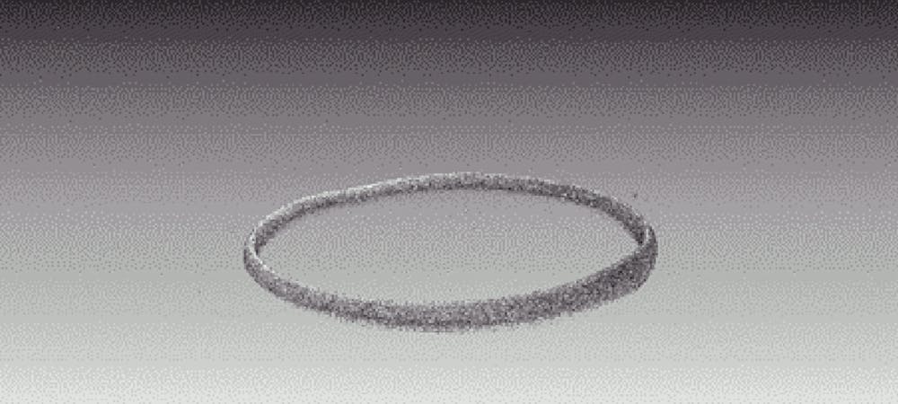
By manipulating the environment the fibroblasts were in, researchers eventually discovered the conditions needed to grow healthy, ring-shaped fibroblast tissues in agarose gel molds. They published these conditions in a study.
This 3D model allows researchers to study the effects of potential drugs to treat fibrosis more accurately than ever before, Morgan said. In 2D models, new drugs to treat fibrosis are evaluated based on their effect on collagen production instead of their direct mechanical effects on the cells.
“Collagen is one of the main proteins for building the extracellular matrix, but there are other proteins that regulate it,” Morgan said. He said that using ECM protein levels to judge the efficacy of a drug is like judging the strength of a house solely on the amount of materials you have to build it.
With this new 3D model, “what we’re able to do, once that structure is made, (is) measure its mechanics, which is the sum of all those parts brought together and built,” Morgan said.
In their most recent publication, the researchers demonstrated how the mechanical attributes of these tissues, such as tensile strength, can be quantified. They used a device called an Instron to stretch the fibroblast rings exposed to pro-fibrotic or anti-fibrotic drugs, which induce and fight off fibrosis, respectively, and measure changes in tissue strain.
This data “is fascinating because we showed that a growth factor like (transforming growth factor) beta decreases the stiffness and tensile strength of (fibroblast rings), and a drug that's anti TGF-beta does the opposite. This shows that we can correlate drug action and growth factor action to tissue mechanics,” Morgan said.
Eric Darling, an associate professor of medical science, engineering and orthopedics who was not involved in the study, wrote that this paper “describes the development of a powerful new approach for evaluating connective tissues in a highly controlled, reproducible way… for their structural, mechanical and biochemical properties,” in an email to the Herald.
Morgan’s lab is continuing to use this model to test the effects of different growth factors and potential fibrosis drugs on connective tissue. He hopes that this model will also be useful for studying genetic connective tissue disorders like Marfan syndrome and Ehlers-Danlos syndrome. In addition, it can be used as a basic science tool to better understand cellular and molecular biology of fibrosis, he said.
“The versatility of the platform is its real strength,” Darling wrote. “The directions that can be explored are nearly endless.”